- -5,00 €


Concise, readable text and an outstanding art program make Gray's Anatomy for Students, 5th Edition, your go-to text for essential information in human anatomy. This fully revised volume focuses on the core information medical students need to know, in an easy-access format and with additional multimedia content to facilitate effective study and mastery of the material. A team of expert authors share a wealth of diverse teaching and clinical experience—all enhanced by more than 1,000 innovative, original illustrations by renowned illustrators Richard Tibbitts and Paul Richardson, who capture anatomical features with unrivalled clarity.
Scheda tecnica
Questa guida alla dissezione, unica nel suo genere, utilizza foto a colori per consentire allo studente di orientarsi più velocemente nel laboratorio di anatomia e sottolinea la rilevanza clinica di ogni struttura e di ogni dissezione.
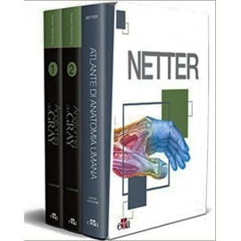
La nuova edizione del GRAY, il volume coordinato da Susan Standring che riscuote in tutto il mondo da 160 anni un successo crescente, è presentata da Edra in abbinamento al NETTER, l'Atlante dall'iconografia inconfondibile. Un'offerta formativa di eccellenza e un'occasione vantaggiosa.
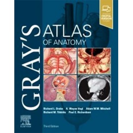
Clinically focused, consistently and clearly illustrated, and logically organized, Gray's Atlas of Anatomy, the companion resource to the popular Gray's Anatomy for Students, presents a vivid, visual depiction of anatomical structures. Stunning illustrations demonstrate the correlation of structures with clinical images and surface anatomy - essential for proper identification in the dissection lab and successful preparation for course exams.
Parte terza: Traumatologia, valutazione clinica e trattamento
Questo libro è indirizzato agli studenti per i quali la conoscenza dell'apparato locomotore rappresenta un sapere fondamentale ai fini della specializzazione professionale. Vengono trattate, come premessa per un corretto approccio allo studio dell'osteoartromiolo...
Originally published as part of the McMinn anatomy atlas family, McMinn's Color Atlas of Head and Neck Anatomy remains the only large format photographic atlas of the human head and neck, incorporating outstanding dissections, osteology, radiographic and surface anatomy images. It is the ideal study aid or trusted reference for the range of students and practitioners who require a detailed understanding of the head and neck, including those in dentistry, radiology and surgery.
For students and clinical professionals who are learning anatomy, participating in a dissection lab, sharing anatomy knowledge with patients, or refreshing their anatomy knowledge, the Netter Atlas of Human Anatomy illustrates the body, region by region, in clear, brilliant detail from a clinician’s perspective. Unique among anatomy atlases, it contains illustrations that emphasize anatomic relationships that are most important to the clinician in training and practice. Illustrated by clinicians, for clinicians, it contains more than 550 exquisite plates plus dozens of carefully selected radiologic images for common views.
Three precious volumes bound in canvas and leather – Size 22 x 28 cm – Over 2500 color illustrations
Fixed-time access to the web platform Virtual Campus and download of the e-books
+
Topographic approach Volume bound in canvas and leather – Size 22 x 28 cm – Over 1000 color illustrations
Fixed-time access to the web platform Virtual Campus and download of the e-book
+
Atlas Volume bound in canvas and leather – Size 22 x 28 cm – Over 1000 color illustrations –
Fixed-time access to the web platform Virtual Campus and download of the e-book
(i cinque volumi sono contenuti in un pratico zaino)
The Netter Atlas of Human Anatomy contains unparalleled, world-renowned illustrations that emphasize anatomic relationships that are most important to the clinician in training and in practice. Unique among anatomy atlases, with illustrations created by clinicians, for clinicians, the fully revised 9th Edition illustrates the body, region by region, in clear, brilliant detail from a clinical perspective, providing more than 550 exquisite plates plus dozens of carefully selected radiologic images for common views. This masterful, foundational content empowers readers with vital anatomy knowledge—whether studying anatomical donor gifts in a dissection lab, visualizing living anatomy through clinical imaging, performing a physical exam, or observing a surgical procedure.
Nervi Cranici
Nervi Spinali
Questo volume di Anatomia umana funzionale comprende la II edizione del testo di Anatomia umana e istologia integrato con un approfondito capitolo dedicato all’Anatomia funzionale dell’Apparato locomotore, allo scopo di offrire allo studente le informazioni necessarie per la conoscenza dell’Anatomia umana e dell’Istologia. Il volume si propone quindi come supporto didattico ideale per gli studenti del corso di Laurea di Scienze Motorie, per quelli di indirizzo sanitario, in particolare i Tecnici Sanitari di Radiologia Medica e i Fisioterapisti, e più in generale per tutti i corsi di Laurea che necessitano di una approfondita conoscenza dell’apparato locomotore.
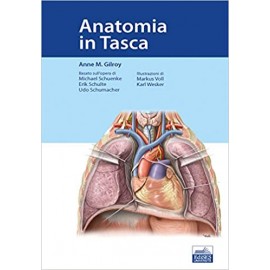
L’Anatomia Umana è una materia che si sviluppa e si apprende soprattutto attraverso le immagini. Questo Atlante tascabile ha lo scopo di rappresentare un valido ed esauriente complemento didattico e un compagno di tutti i giorni per l’apprendimento dell’Anatomia Umana in tutti i Corsi di Laurea a indirizzo Sanitario e in quello di Scienze Motorie.
Il suo formato tascabile ne permette una rapida consultazione che potrà rivelarsi utile anche in numerose occasioni della quotidiana attività clinica e professionale.