- -10,00 €
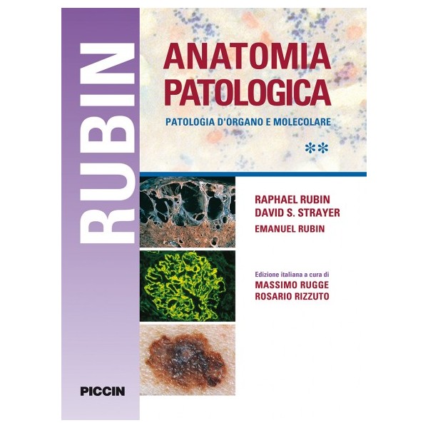





Edizione italiana a cura di Massimo Rugge e Rosario Rizzuto.
Volume riccamente illustrato a colori di 1050 pagine
Scheda tecnica
This expert volume in the Diagnostic Pathology series is an excellent point-of-care resource for practitioners at all levels of experience and training. Covering the full range of nonneoplastic and neoplastic conditions of the female genital tract, it incorporates the most recent scientific and technical knowledge in the field to provide a comprehensive overview of all key issues relevant to today’s practice. Richly illustrated and easy to use, the third edition of Diagnostic Pathology: Gynecological is a visually stunning, one-stop resource for every practicing pathologist, resident, student, or fellow as an ideal day-to-day reference or as a reliable training resource.
A Volume in the Pattern Recognition Series
Part of the in-depth and practical Pattern Recognition series, Practical Surgical Soft Tissue Pathology, 2nd Edition, helps you arrive at an accurate diagnosis by using a proven pattern-based approach. Leading diagnosticians guide you through the most common patterns seen in soft tissue pathology, applying appropriate immunohistochemistry and molecular testing, avoiding pitfalls, and making the best diagnosis. High-quality illustrations capture key morphologic patterns for a full range of common and rare tumor types, and a "visual index" at the beginning of the book directs you to the exact location of in-depth diagnostic guidance.
For over 60 years, residents and practicing pathologists have turned to Rosai and Ackerman's Surgical Pathology for definitive guidance on every aspect of the field, delivered in a readable, easy-to-digest, and engaging manner. In the two-volume 11th Edition, a dynamic new author team ensures that this classic text retains its signature anecdotal style, while revising the content to bring you fully up to date. Widely used for board exam preparation, as well as for everyday reference in practice, this leading resource equips you to effectively and efficiently diagnose the complete range of neoplastic and non-neoplastic entities.
AFIP ATLAS OF TUMORS AND NON-TOMORS PATHOLOGY. SERIE 5
With its consistent, practical organization and succinct bulleted format, Atlas of Head and Neck Pathology, 4th Edition, is an ideal resource for pathologists in training and in practice. High-quality illustrations with extensive figure legends depict the histological, immunohistochemical, cytologic and diagnostic imaging appearances of every type of head and neck pathology, while the user-friendly format allows for quick access to information. From cover to cover, this comprehensive reference is designed to help you effectively and accurately diagnose a wide range of head and neck disorders, improve your turnaround time when diagnosing a specimen, and facilitate clear reports on prognosis and therapeutic management options to surgical and medical colleagues.
Contenuti
✔✔Oltre 1500 immagini
✔✔Ciascuna figura è affiancata da un testo sintetico che descrive i punti chiave illustrati
✔✔Comprende esempi di organi e tessuti sia normali sia patologici